
Your cart is currently empty!
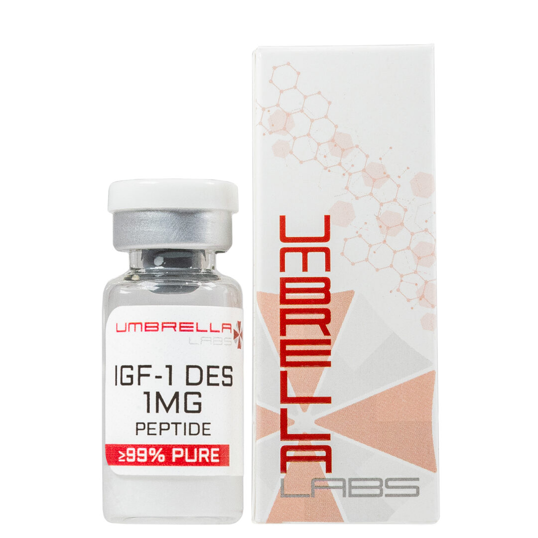
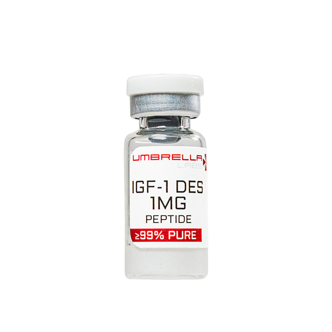
DES IGF-1 – 1 mg
DES (1-3) IGF-1 is modified, more efficient and improved version of the hormone, IGF-1, with approximately 10 times stronger action and effect as the classical IGF-1! DES (1-3) IGF-1 is particularly suited for massive muscle building and using chiefly shortly before and after training.
Description
DES (1-3) IGF-1 is modified, more efficient and improved version of the hormone, IGF-1, with approximately 10 times stronger action and effect as the classical IGF-1! DES (1-3) IGF-1 is particularly suited for massive muscle building and using chiefly shortly before and after training.
It is no secret that IGF-1 is produced in the human body in the liver and in part also in other tissues, but the natural concentration of this hormone is insufficient for achieving the desired results in a significant increase in muscle mass. DES (1-3) IGF-1 is a new generation of IGF-1, which shifts the level and efficiency considerably further. His popularity among athletes and enthusiasts who want as quickly as possible to gain muscle mass because of sharply increasing day by day.
The effectiveness of DES (1-3) IGF-1 has no doubt athletes talk about excellent results. DES (1-3) IGF-1 has a very strong anabolic effects – stimulates the metabolism of amino acids in the body (increases protein synthesis) increases RNA synthesis, promotes fat metabolism, glucose transport and has a strong anti-catabolic effect (protects muscles from decomposition). IGF-1 can stimulate hyperplasia – the growth of new cells. Moreover, the ability to bind to receptors of lactic acid, and enables to start the process of formation of new muscle tissue directly during training and not only afterwards.
DES (1-3) IGF-1 has a very low affinity for binding proteins only 1%, which makes it very effectively usable form of IGF-1, while up to 98% of standard IGF-1 bound to the binding proteins (and remains inactive It is not available for use on skeletal muscles and stimulate its growth). That is the main reason for his secrecy and high efficiency.
Effects DES (1-3) IGF-1:
- DES (1-3) IGF-1 has a very strong anabolic effects (increased protein synthesis, and building a massive muscle)
- DES (1-3) IGF-1 has a strong anti-catabolic effect
- DES (1-3) IGF-1 induces hyperplasia – the growth of new muscle cells!
- DES (1-3) IGF-1 is not detectable during doping controls
- DES (1-3) IGF-1 promotes regeneration of fast and strong
- DES (1-3) IGF-1 promotes increased immunity and regeneration of the nervous system
Differences between DES (1-3) IGF-1 and IGF-1 LR3:
- – Disadvantage DES (1-3) IGF-1 is short effect – the half-life is approximately 20 minutes, while IGF-1 LR3 are substantially longer – after about 24 hours
- + Advantage DES (1-3) is approx. up to 5 times stronger anabolic effect as that of IGF-1 LR3
- + Due to the short lifetime of the DES treated with (1-3) IGF-1 is preferably mainly in the local muscles to which it has been applied (which can be quite valuable feature when trying to develop or improve certain preferred (e.g. lagging muscle groups), IGF -1 LR3 act more globally, or the entire body evenly on all muscle groups
- – Disadvantage DES (1-3) IGF-1, more frequent – several times daily, for IGF-1 LR3 sufficient dose 1 day
- – DES (1-3) IGF-1 may cause at high doses hypoglycaemia
At the recommended dose DES (1-3) IGF-1 has no side effects. The recommended dose for one injection is from 20 to 50 mcg of the DES (1-3) IGF-1 to muscle intramuscularly. The dosing frequency is typically 3-5 times per day, although DES (1-3) IGF-1 may be due to the short life theoretically administered every 20 minutes, if desired (which is very practical terms almost unreal, financially) .As most important thing we could mark a dose of approx. 5 minutes before your workout (which ensure the start of the process of creating new muscle and tissue directly during training), and other benefits after training. As regards the duration of treatment with DES (1-3) IGF-1 may be selected individually and ranges from 4 to 12 weeks. It is recommended to use a break in about 5 weeks.
The second method of administration involves the combined use of PEG-MGF & DES IGF-1 with each other in a common interaction (at which bind to each other processes of proliferation and differentiation), in order to optimize the resulting hyperplasia target local muscle. With this method, a first PEG-MGF administered separately the first 1-4 weeks, then followed by administration of DES IGF-1, for the same number of weeks. Here it should be noted that scientists and athletes are still just learning and exploring how this optimal drug use in this case.
- IGF-1 DES is a truncated variant of insulin-like growth factor-1 (IGF-1). IGF-1 is typically composed of 70 amino acids but IGF-1 DES is a shortened analogue with only 67 amino acids. The first three amino acids that are emitted are Gly-Pro-Glu which are removed through the N-terminus. By shortening the peptide chain IGF-1 becomes 10 times more potent in terms of increasing hypertrophy as well as reducing the binding of IGF-1 binding proteins due to the absence of glutamate.
- Increasing the potency is important when it comes to the efficacy of the peptide and maximizing muscle growth. The improved potency comes from the ability of IGF-1 DES to efficiently bind to lactic acid receptors. IGF-1 DES is capable of binding to certain cellular receptors that have been mutated due to the presence of lactic acid, allowing for increased signaling for tissue growth. However, this truncated form of the peptide has a short half-life of only about 20-30 minutes [1].
- Main Research Findings
- 1) Results of this study reported that IGF-1 DES led to excitatory transmission in the CA1 region of the rat hippocampus and enhancement of fEPSP slopes in a dose-dependent manner.
- 2) The research team of Elis et. Al found in KID and KIR mice models, IGF-1 is crucial for the development of normal body, organ, and bone size.
- 3) Results of the study found that subcutaneous injection of IGF-1 DES at the onset of diabetes can act as a protective measure against predegenerative biochemical abnormalities.
- Selected Data
- 1) The research team utilized male Sprauge-Dawley rats as test subjects for this experiment. Once the subjects reached 20-40 days old they were anesthetized with halothane in order for researchers to obtain slices of certain brain regions. Coronal hippocampal slices were prepared with a vibrating tissue slicer and maintained at room temperature in a solution of oxygenated artificial cerebrospinal fluid (ACSF). The ACSF contained 124mM NaCl, 3.3 mM KCl, 2.4 mM MgCl2, 2.5 mM CaCl2, 1.2 mM KH2PO4, 10 mM D-glucose, and 25.9 mM NaHCO3. The slices were saturated with the ACSF twice per minute during the observation period [2].
- Researchers used NMDA receptor antagonists (APV), GABA receptor antagonists (BIC), and AMPA/KA receptor antagonists (DNQX) in order to pharmacologically isolate and observe evoked currents. IGF-1 DES was prepared as a stock solution in 0.1 N acetic acid. Various compounds were used in order to examine the signaling components of IGF-1, including, tyrosine kinase inhibitors, PI3K inhibitors and LY 294002 inhibitors. Like IGF-1 DES these inhibitors were made into a stock solution, however, instead of acetic acid dimethyl sulfoxide (DMSO) was used. All drugs were administered via ACSF [2].
- In order to collect data electrodes were prepared and filled with a recording solution containing 120 mM K-gluconate, 10 mM KCl2, 5 N-(2,6-dimethyl-phenylcarbamoylmethyl)-triethylammonium bromide, 1mM EGTA, 0.1 mM CaCl2, 2 Mg-ATP, 0.2 tris-GTP, and 10 mM free acid (HEPES). Neuron voltages were clamped at -70 mV in order to record AMPA-mediated excitatory postsynaptic currents (EPSCs) while in the presence of antagonists, APV and BIC. Neuron voltages were clamped and -30 mV in order to record NMDA-mediated EPSCs while in the presence of DNQX and BIC. The synaptic currents were sent every 20 seconds by way of a 0.2 ms long electrical stimulation of the adjacent tissue to the electrode [2].
- Field excitatory postsynaptic potentials (fEPSPs) were obtained under the same conditions and using the same recording equipment as the patch-clamp recordings. The only change made was the adjustment of the recording electrodes which were now placed in the apical dendritic field. The IGF-1 DES-induced concentration-response curves of EPSCs and fEPSPs were analyzed by one-way ANOVA and Newman-Keuls test for comparisons. Dunnett’s post hoc test and one-way ANOVA tests were used to compare inhibitory effects on IGF-1 DES-mediated changes in fEPSP slopes [2].
- 2) The study conducted by Elis et. Al aimed to find how bioavailable derivatives of IGF-1 affects somatic and skeletal growth as well as body composition and tissue integrity. The research team created two mutated mouse models with knock-in RE-IGF1 and IGF-1 DES. The first mutant substituted an E amino acid for an R at position three and was named knock-in R (KIR). While the second mutant is missing the first three amino acids of IGF-1 (IGF-1 DES) and was named knock-in D (KID). The two mutated mouse models represent an unbound form of IGF-1 with impaired post-translational control and increased bioavailability; they are then used to characterize the effects of KIR and KID in this scenario [3].
- Serum levels of IGF-1 in KID and KIR mice were observed by using a radioimmunoassay (RIA) kit. The same RIA kit was used to measure recombinant RE-IGF1 and IGF-1 DES. The bioactivity of free IGF1 in serum was observed using a cell-based IGF-1 kinase receptor activation (KIRA) bioassay. This is used to determine the ability of the serum to stimulate IGF1R activity in vitro [3].
- 3) 12-week-old male Sprague-Dawley rats were assigned to different treatment groups and used for animal experimentation in accordance with NIH guidelines. The animals were split into two experimental groups and a control group, each group had 5 rats. All solutions were administered to the rats through Acrodisk filters. The experimental groups were intraperitoneally administered a 50 mg/kg dose of STZ in order to induce diabetes. The control group was labeled non-diabetic (ND) while the first experimental group was labeled at STZ-veh indicating the animal was diabetic and received vehicle treatments. The second experimental group was labeled STZ-des, the diabetic rats received an active dose of IGF-1 DES. Each treatment was administered through a subcutaneous osmotic minipump over the course of two weeks [4].
- After two weeks of treatment the rats were euthanized and the eyes were preserved with 4% paraformaldehyde in phosphate buffered saline (PBS). The preserved eyes were set in paraffin and cut into slices measuring approximately 4 micrometers in length. Additionally, tail blood was collected in order to measure glucose assay 24 hours after STZ or vehicle treatment and again at 2 weeks after treatment. The retinal tissue slices were rehydrated through the use of xylene and various graded alcohol concentrations. The samples were then properly prepared by rinsing in PBS and incubation with the primary antibodies. The slices were stained in order to identify the number of immunoreactive cells in the ganglion cell layer (GCL) and the inner nuclear layer (INL) throughout three randomly chosen segments [4].


Reviews
There are no reviews yet.