
Your cart is currently empty!
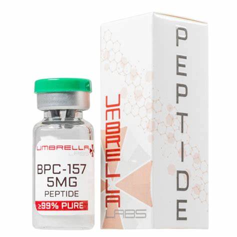
BPC-157 – 5 mg
BPC-157 (Body Protection Compound-157) is a pentadecapeptide made up of 15 amino acids. The sequence of amino acids in BPC-157 is similar to the amino acid sequence in a portion of human BPC.
Description
BPC-157 (Body Protection Compound-157) is a pentadecapeptide made up of 15 amino acids. The sequence of amino acids in BPC-157 is similar to the amino acid sequence in a portion of human BPC. Human BPC is found in gastric juice. Experiments have shown that BPC-157 promotes wound healing, including tendon injuries such as Achilles tendon rupture in rats. The aim of this study was to investigate the likely mechanism that BPC-157 uses to accelerate the healing process in the injured tendon. The study used two groups of tendon explants. One group was cultured in medium containing BPC-157 and the other in medium without BPC-157. Tendon fibroblast outgrowths were then observed in these cultures. Such outgrowths indicated tendon regeneration. The results revealed that the growth of the explant was significantly accelerated in the culture containing BPC-157 compared to the culture without BPC-157. In addition, MTT assay showed that BPC-157 had no direct effect on cell proliferation in the culture derived from rat Achilles tendon. However, the results also showed that BPC-157 significantly increased cell survival under oxidative stress. In addition, the Transwell filter migration assay showed that BPC-157 significantly increased fibroblast migration in vitro in a dose-dependent manner. BPC-157 also increased the dispersion of fibroblasts in culture dishes in a dose-dependent manner.
✅the effective combination for maximum recovery after injury is BPC-157+TB-500.
BPC 157 is characterized as a pentadecapeptide containing partial sequence of the body protection compound (BPC) that is shown to have had positive effects on the efficiency and efficacy of various growth hormones as well as various other healing qualities in cases such as spinal cord injury and burn wounds.
3 Main Research Findings
1) Application of BPC 157 improved the burn-wound healing ability of mice while attenuating burn-gastric lesions.
2) Administration of the peptide BPC 157 enhances the expression of growth hormone receptors in tendon fibroblasts.
3) Evidence shows that administering BPC 157 to rats improves the healing rate of spinal cord injury while enhancing functional recovery.
Selected Data
1) The research team of Mikus et. Al examined the effects of topical application and intraperitoneal application of the BPC 157 on burn-wound healing burn-gastric lesions. The peptide was acquired as a 99% pure substance that was further dissolved in saline prior to the random assignment of male mice to an experimental treatment. All subjects were housed in individual cages with ad libitum access to food and water. Each experimental treatment group included 10 mice. While sedated under ether anesthesia, deep partial skin thickness burns were induced on the back of each test subject through the method of control burning. The first application of the peptide was given immediately after the burn-injury. 50 ug of BPC 157 was either dissolved in 2 ml of distilled water or mixed with 50 g of a commercial neutral cream to be applied in a line layer over the burn. Similarly, 10 ug or 10ng/kg of BPC 157 was also administered to a group of test subjects through an intraperitoneal injection. Additionally, an equal amount of neutral commercial cream or saline was topically and intraperitoneally administered to the test subjects in order to act as a vehicle-control [1].
The burn healing parameter was evaluated through various forms of histological investigation, water content assessment in the burned skin, and tensiometry studies such as breaking strength and elongation of the burned skin. The same methods were used to assess the status of the induced gastric lesions; all assessments were completed on either day 1, 2, 3, 7, 14, or 21. The histological assessment used to investigate the healing parameters of the mice included edema, formation of blood vessels, retained follicles, formation of reticulin and collagen, number of inflammatory cells, and the number of subjects experiencing complete re-epithelialization. The healing parameters were tested on different days following euthanization of the animals. The schedule is as follows: day 1: edema, diameter and number of blood vessels, number of inflammatory cells and reticulin, and number of vital follicles; day 2 and 3: day 1 necrosis; day 14: number and diameter of blood vessels, reticulin and collagen levels, epithelization; day 21; number and diameter of blood vessels, epithelization and collagen.
When assessing the water content of the burned sites, the researchers initially began by conducting a separate experiment that included a 20×20 mm specimen of full-thickness tissue originating from the burned sites of the back. Additionally, a 20×20 mm specimen was extracted from the non-damaged tissue of the upper back for the purpose of weight determination at days 1, 2, and 3 following euthanization. All tissue biopsies were placed in a drying chamber until a stable weight was measured for approximately 48 hours. The research team then calculated the wet/dry ratio of each specimen in order to compare them to the values of the control groups. Tensiometry investigation took place by measuring the tensile breaking force and relative elongation of the damaged skin immediately after euthanasia and at days 7 and 14. A special device obtained from the Technical Faculty at the University of Zagreb, Croatia using the formulas: breaking force (N/mm2)=force (N)/cross-section skin burned area (mm2); and relative elongation of burned part of skin=Δlength burned skin immediately before breaking/initial length of burned skin. This protocol was followed by statistical analysis completed by Fisher’s exact probability test two-tailed and one-way ANOVA [1].
2) The research team of Chang et. Al examined the effects of BPC 157 on the expression of growth hormones and how it can potentially activate fibroblast proliferation and keratinocyte migration. Growth hormones are peptides that stimulate growth, cell reproduction, and regeneration of chondrocytes and osteoblasts, as well as increase protein production and muscle mass. The study utilized male Sprague-Dawley rats as their source of tendon fibroblasts; the achilles tendons were harvested by aseptic procedures followed by dissection into pieces at size about 1.5 to 2.0 mm^3 and separation into six-well culture plates. After migrating out from the explants, the tendon fibroblasts began to grow rapidly. Once the samples reached confluency the cells were subcultured and all tendon fibroblasts between passages 2 and 4 that had a proper growth rate and normal fibroblast shape were used in the experimentation process. From there, BPC 157 synthesized and purchased from Kelowna International Scientific Inc. was added to the fibroblasts at concentrations of ) (control group), 0.1, 0.25, and 0.5 ug/mL and incubated in a humidified atmosphere for 1, 2, and 3, days [2].
The next experimental step included extracting total RNA from the cells through the use of the acid guanidinium thiocyanate-phenol-chloroform extraction method. Total RNA was then used to synthesize complementary DNA, followed by the performance of real-time PCR using an SYBR Green I technology and MxPro-Mx3000P QPCR machine. All real-time PCRs were performed three times and all changes in gene expression were reported as multiples of increases relative to the control while GAPDH was used as an internal control and relative gene expression between the experimental groups were determined using MxPro software.
Furthermore, western blot analysis took place by initially preparing cell extracts in a lysis buffer followed by the standard method of ultrasonication. The Bradford assay was used to determine protein concentration of cell extracts, from there samples with the same amount of protein were separated using SDS-PAGE technology and transferred onto a PVDF membrane. The membrane was then incubated at room temperature in blocking solution for 1 hour, followed by incubation in blocking solution containing the appropriate dilution of either phospho-Jak2 or the primary antibody for growth hormone receptor, for 2 hours. The membrane was washed three times in PBS prior to 1 hour of incubation in PBS containing goat anti-mouse IgG conjugated with horseradish peroxidase. After an additional three washings with PBS, positive signals were developed with an enhanced chemiluminescence kit [2].
An MTT assay was performed next by seeding tendon fibroblast in each well of 24-well culture plates. In addition to a medium made of 0.5 ml of DMEM, 10% FBS, 100 U/mL of penicillin, and 100 mg/mL of streptomycin, BPC 157 was added to each well in concentrations of 0 (control group), 0.1, 0.25, and 0.5 ug/mL for 1, 2, and 3 days in a humidified atmosphere. One day after growth hormone was added to the tendon fibroblasts treated with BPC 157, the cells were washed with PBS followed by the addition of 1 mL DMEM containing 0.05 mg/mL MTT,, and incubation for 1 hour. The media was then removed and the formazan crystals that formed during incubation were dissolved in 1 mL DMSO and processed for OD reading.
3) The research team of Pervoic et. Al studied the effectiveness of BPC 157 treatment when healing spinal cord injuries and how the peptide promotes functional recovery in test subjects. This study included 12 week-old Wistar albino male rats that were bred in-house at the animal facility at the Department of Pharmacology, School of Medicine in Zagreb, Croatia. The animals were allowed to acclimate to their new environment over the course of 5 days, followed by random assignment to an experimental treatment group. At least 6 animals were included in each group and all test subjects were maintained under standard conditions including a 12 hour light/12 hour dark schedule as well as ad libitum access to a standard GLP diet and fresh water. BPC 157 was dissolved in 0.9% NaCl and underwent further preparation with 99% high-pressure liquid chromatography (HPLC).
The animals were then deeply anesthetized with 3% isoflurane and 50 mg/kg of ketamine followed by a laminectomy at lumbar level L2-L3, corresponding to the sacrocaudal spinal cord. A 200 ug/kg or 2 ug/kg dose of BPC 157 was intraperitoneally injected 10 minutes post-injury followed by the return of animals to their cages and assignment to interval groups varying from days 7, 15, 20, 90, 180, and 360. The tail motor function of each test subject was scored 8 hours and 1, 4, 7, 15, 30, 90, 180, and 360 days post-injury and measured on a scale of 0-5. 0 was defined as autotomy; 1-complete loss of tail function; 2-maximum elevation of ¼ of the tail; 3-maximum elevation of ½ of the tail; 4-maximum elevation of ¾ of the tail; and 5 was defined as normal functioning of tail motor control. The same intervals were used when assessing tail spasticity. Manual stimulation of the tails was performed next using the standardized stretch/rub maneuver followed by scoring according to the Bennett scale. 0 on the Bennett scale is defined as a normal phenotype; 1-flaccid tail; 2-hypertonic flexor muscle, tail coiled and stiff; 3- hyperreflexia; and 4 is defined as hypertonic flexor and extensor muscles [3].
Prior to euthanization, the animals from the 30, 90, 190, and 360 day post-injury interval groups were placed in a wooden box that allowed for tail exposure. This was followed by three pairs of monopolar needles stabbed 3 mm deep into the tail at measurements of 10, 60, and 100 mm caudal to the base of the tail. From there the voluntary muscle activity was recorded from the most caudal pair of electrodes, followed by measurement and recording of the average motor unit potential (UMP) and the compound motor action potential (CMAP). CMAP was recorded after stimulating the first and second electrodes followed by recording of the amplitude, proximal and distal latencies, and polyphasic changes. Following the electrophysiological testing, histology testing took place by initially collecting a 10-mm long piece of the spinal column and the surrounding muscle. After 5 um cross-sections were prepared according to procedure, the intensity and distribution of the pathological spinal cord changes were evaluated semiquantitatively. In this study 0 was defined as no changes; 1-small or focal changes; 2-moderate changes; and 3-numerous confluent changes subcategorized into: (a) the hemorrhagic zone, (b) edema, (c) the loss of neurons, (d) vacuoles, and (e) the loss of spinal column tracts [3].
| CAS Number | 137525-51-0 |
| Other Names | BPC157, BPC 157, Bepecin, 8ED8NXK95P, Bpc 15, PLD-116, PL-14736, pl-10, Booly protection compound 15 |
| IUPAC Name | (4S)-4-[(2-aminoacetyl)amino]-5-[(2S)-2-[(2S)-2-[(2S)-2-[[2-[[(2S)-6-amino-1-[(2S)-2-[[(2S)-1-[[(2S)-3-carboxy-1-[[(2S)-3-carboxy-1-[[(2S)-1-[[2-[[(2S)-1-[[(1S)-1-carboxy-2-methylpropyl]amino]-4-methyl-1-oxopentan-2-yl]amino]-2-oxoethyl]amino]-1-oxopropan-2-yl]amino]-1-oxopropan-2-yl]amino]-1-oxopropan-2-yl]amino]-1-oxopropan-2-yl]carbamoyl]pyrrolidin-1-yl]-1-oxohexan-2-yl]amino]-2-oxoethyl]carbamoyl]pyrrolidine-1-carbonyl]pyrrolidine-1-carbonyl]pyrrolidin-1-yl]-5-oxopentanoic acid |
| Molecular Formula | C₆₂H₉₈N₁₆O₂₂ |


Reviews
There are no reviews yet.